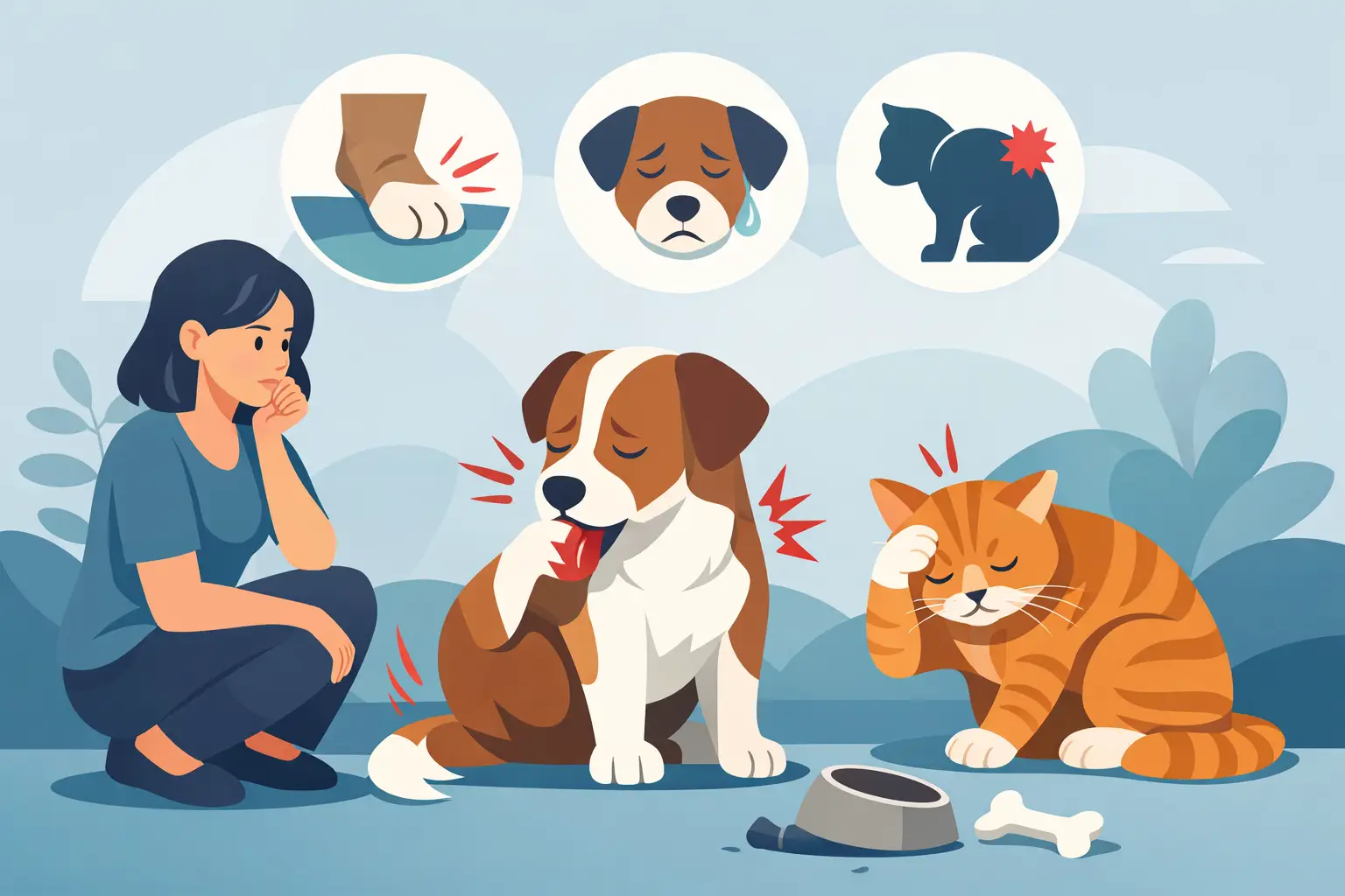
When your dog suddenly starts limping or your cat stops jumping onto the couch, answers matter fast. Digital radigraphs give veterinarians a quick, detailed way to look beneath the surface and find out what may be causing pain, illness, or changes in movement.
For many pet owners, X-rays feel familiar in theory but less clear in practice. You may know your pet needs imaging, but not what the test shows, how long it takes, or whether it will be stressful for your animal. That uncertainty is common. The good news is that digital imaging has made the process faster, clearer, and more useful in everyday veterinary care.
What digital radigraphs are
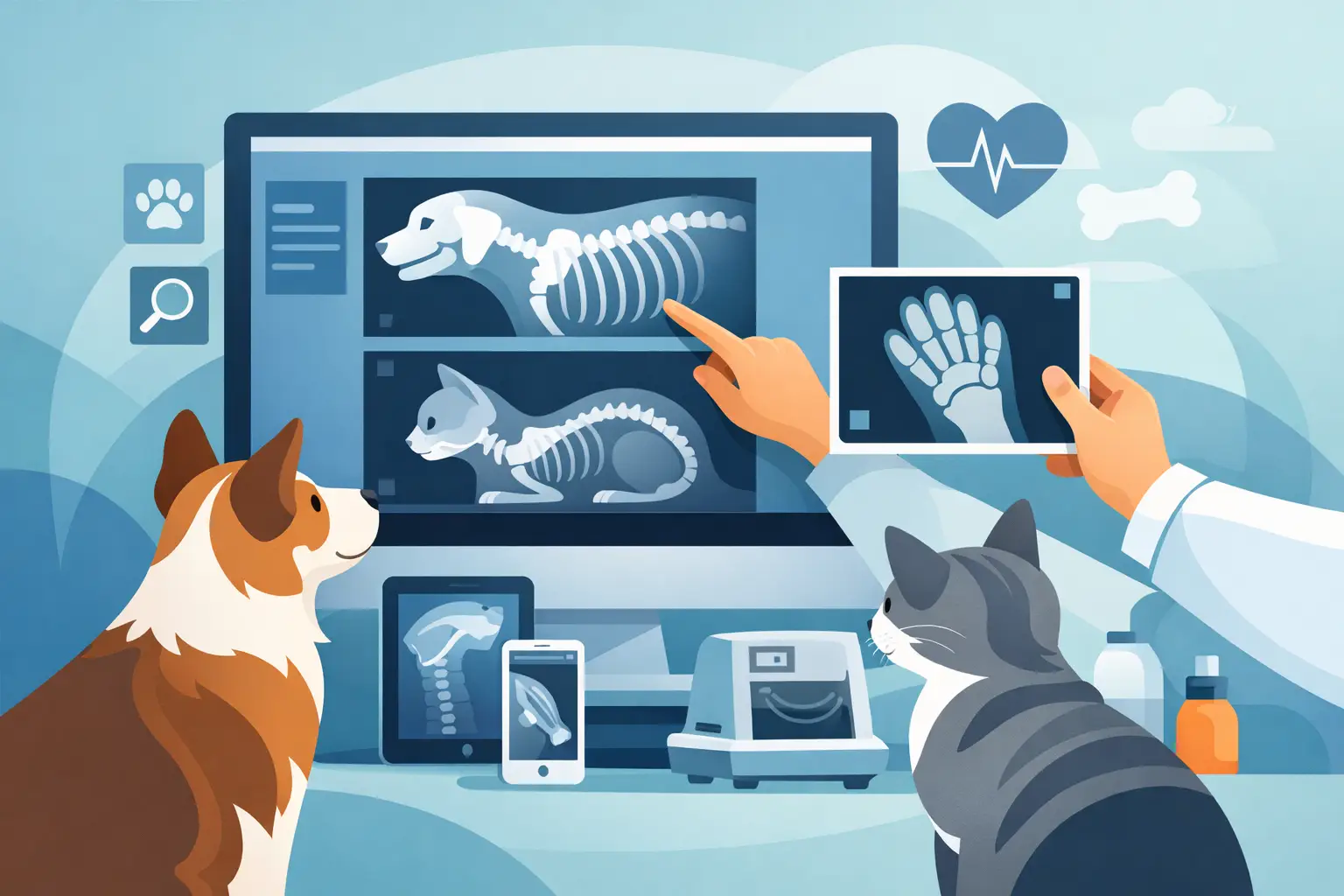
Digital radigraphs are veterinary X-rays captured electronically instead of on traditional film. They create images of your pet’s bones, chest, abdomen, teeth, and other internal structures, allowing your veterinary team to evaluate problems that cannot be confirmed with a physical exam alone.
The word itself is often used interchangeably with digital X-rays. In a clinical setting, the goal is straightforward: get a clear picture quickly so your veterinarian can make an informed recommendation. That may mean confirming a fracture, checking for arthritis, evaluating the lungs, looking for bladder stones, or assessing the abdomen when a pet is vomiting or painful.
Because the images are digital, they can usually be viewed within minutes. That speed matters when a pet is uncomfortable, when an injury needs prompt treatment, or when your veterinarian is deciding whether additional testing is needed.
Why veterinarians use digital radigraphs
Pets cannot explain where it hurts, when a symptom started, or whether they swallowed something they should not have. Imaging helps fill in those gaps. A careful exam still comes first, but some conditions only become visible when your veterinarian can see what is happening internally.
Digital radigraphs are especially helpful for orthopedic concerns. If your dog is favoring a leg, has stiffness after rest, or seems reluctant to climb stairs, X-rays can help identify fractures, joint changes, hip issues, or signs of degenerative disease. In older pets, they may reveal arthritis that has been building gradually over time.
They are also valuable for chest and abdominal cases. A cough may call for chest imaging to look at the lungs and heart. Vomiting, decreased appetite, or abdominal discomfort may lead your veterinarian to evaluate the stomach, intestines, or bladder. Sometimes the images provide a clear answer right away. Other times, they help rule out major concerns and guide the next step, such as ultrasound or lab work.
Dental care is another important area. Many painful dental problems sit below the gumline, where they are impossible to judge fully by sight alone. Digital dental radiographs can show tooth root infection, bone loss, retained roots, and other hidden issues that affect comfort and treatment planning.
How digital radigraphs differ from traditional X-rays
The biggest difference is efficiency. Traditional film X-rays require processing time and offer less flexibility once the image is taken. Digital systems produce images much faster and allow the veterinary team to enlarge, adjust contrast, and review details more easily.
That does not mean every case becomes simple. Positioning still matters, and some pets need multiple views so the area can be assessed accurately. But digital imaging often shortens the overall process and improves image quality, which supports faster decisions.
There is also a practical benefit for pet owners. Clearer images can make conversations easier. When your veterinarian can show you what they are seeing, whether it is a broken bone, spinal change, enlarged organ, or swallowed object, treatment recommendations tend to feel more understandable and less abstract.
What to expect during your pet’s appointment
In many cases, digital radigraphs are a routine and well-tolerated part of a visit. Your veterinary team will start by examining your pet and asking questions about symptoms, activity changes, appetite, breathing, or recent injuries. If imaging is recommended, they will explain what they are looking for and why.
Your pet then needs to be positioned carefully so the images are useful. That may sound simple, but pets do not naturally hold still in the exact position needed for a diagnostic image. Some animals remain calm with gentle handling and reassurance. Others are painful, anxious, or too wiggly to position safely without extra help.
That is where the plan becomes individualized. Some pets can have digital radigraphs while fully awake. Others benefit from light sedation, especially if the area being examined is painful or if precise positioning is essential. Sedation is not automatically required, and it is not a sign that something is wrong. It is simply one tool veterinarians use to reduce stress, improve safety, and obtain accurate images.
When sedation may be recommended
Pet owners sometimes worry when sedation comes up, but there are good reasons it may be the best choice. A frightened dog with an injured leg may tense up or resist movement. A painful cat may become defensive when handled. If your pet is stressed, struggling, or uncomfortable, trying to force the process can increase fear and may still result in poor-quality images.
Light sedation can make the experience smoother for everyone involved. It can also prevent the need for repeat images, which saves time and can reduce overall stress. The trade-off is that sedation adds another layer of planning, and your veterinarian will consider your pet’s age, medical history, current condition, and the urgency of the situation before recommending it.
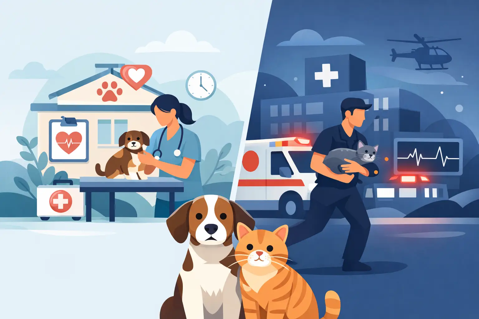
For some emergencies, imaging must happen quickly and the approach is adapted to the pet’s stability. For elective or non-urgent concerns, your veterinarian may suggest timing, pre-sedation instructions, or additional testing first. It depends on the case.
What digital radigraphs can and cannot show
Digital radigraphs are extremely useful, but they are not the answer to every diagnostic question. They are excellent for evaluating bones, many chest conditions, certain abdominal problems, and dental structures. They can suggest when something is abnormal and help narrow down possibilities.
At the same time, some soft tissue problems do not show up clearly on X-rays alone. A normal radiograph does not always mean nothing is wrong. Early disease, small masses, ligament injuries, and some gastrointestinal issues may require other diagnostics for a fuller picture.
That is why veterinary medicine works best as a combination of tools. A physical exam, medical history, lab work, digital radigraphs, ultrasound, and sometimes advanced imaging all play different roles. The right next step depends on what your pet is showing and what the initial findings reveal.
Why speed and clarity matter in a full-service hospital
When imaging is available as part of a broader in-house care approach, decisions can happen more efficiently. If your pet comes in for limping, coughing, trauma, or abdominal pain, digital radigraphs can often be performed as part of the same workup rather than delayed or referred elsewhere.
That continuity matters for families who want answers without bouncing between multiple locations. It also helps the veterinary team connect the dots. Imaging results can be reviewed alongside the physical exam, bloodwork, surgery needs, rehabilitation goals, or dental findings to build a treatment plan that makes sense for the whole pet, not just one symptom.
At All Creatures Veterinary Center, that kind of integrated care is part of the goal: helping pets receive routine, urgent, and advanced support in one familiar setting whenever possible.
Questions to ask if your pet needs imaging
If your veterinarian recommends digital radigraphs, it is reasonable to ask what they are looking for, whether sedation is likely, how soon results will be reviewed, and what the next step may be depending on the findings. Good veterinary care includes clear communication, not just technology.
You can also ask how the imaging fits into the bigger plan. Sometimes the goal is to confirm a likely diagnosis. Sometimes it is to rule out dangerous possibilities. And sometimes it is the first piece of a larger diagnostic process. Knowing which situation applies can help you feel more prepared.
A recommendation for imaging does not always mean the worst. Often, it is the most practical way to replace uncertainty with useful information.
When your pet is hurting or not acting like themselves, waiting and wondering can be the hardest part. Clear imaging, thoughtful interpretation, and a veterinary team that takes time to explain the findings can make the next decision feel a little less overwhelming.