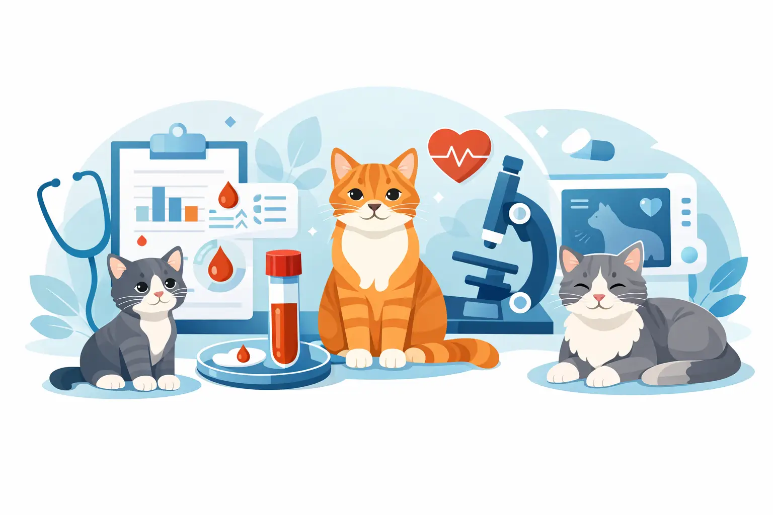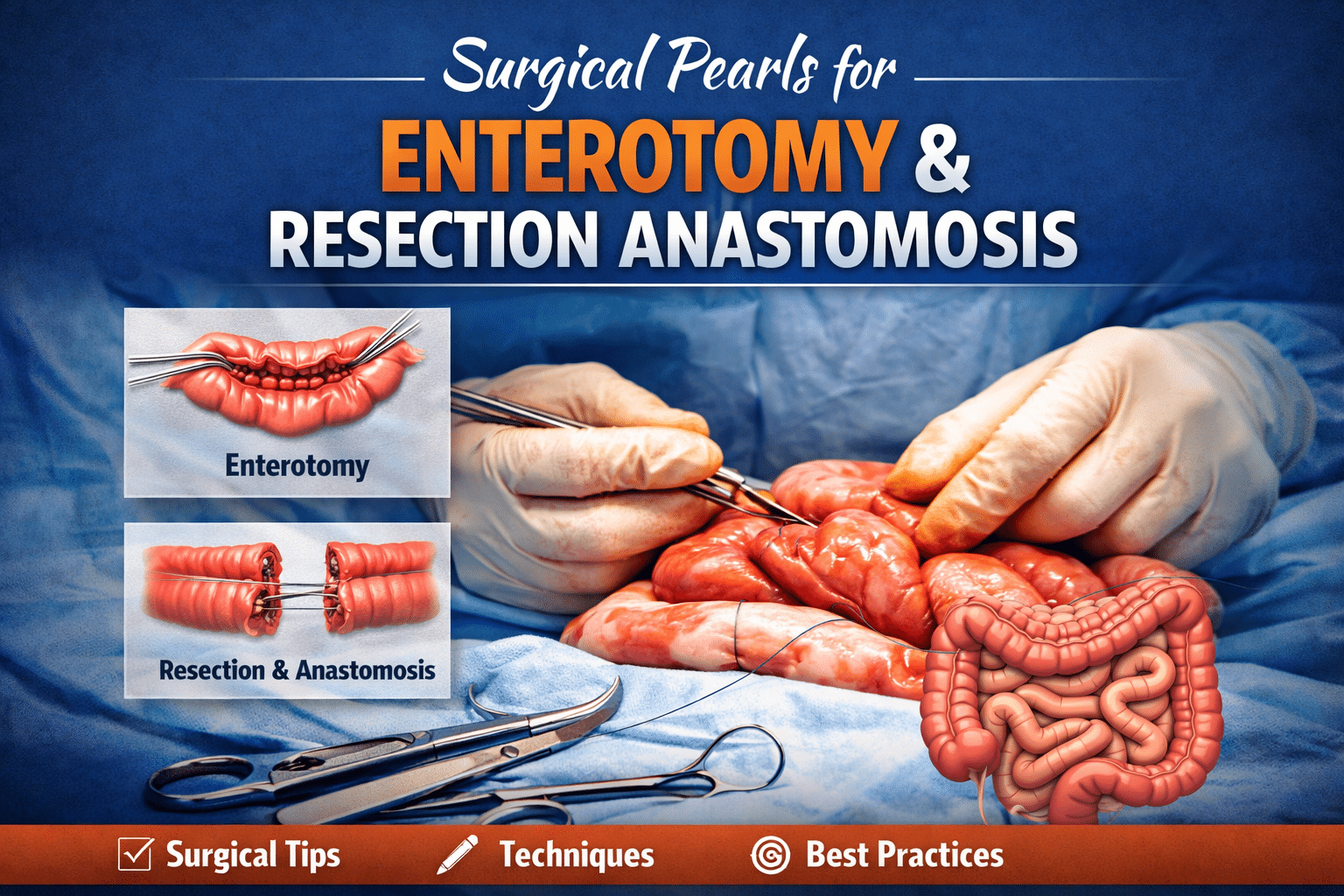
Surgical Pearls for Enterotomy and Resection & Anastomosis
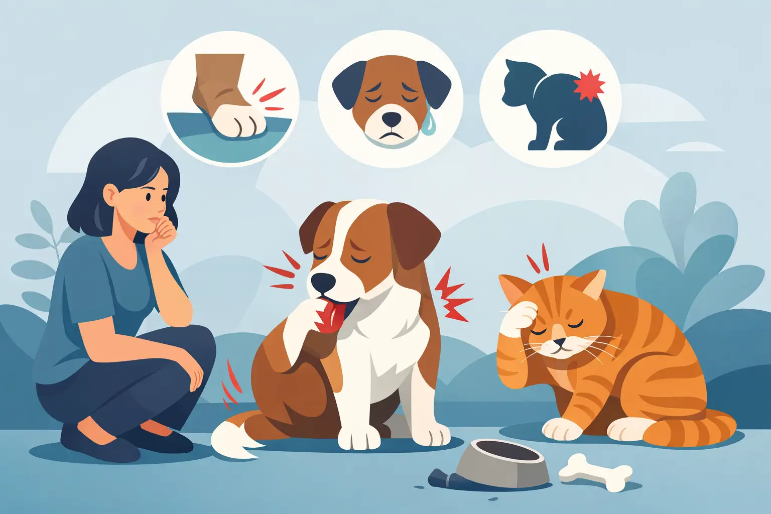
Surgical procedures for the intestines are pretty common in pets. Enterotomy is a surgery done on the pet’s intestine to remove the object from the intestine. It is done to remove any unwanted objects that are stuck in the intestine. Resection and anastomosis are the removal of the damaged or diseased part in the pet’s intestine and then stitching the area to let the intestine work in its natural manner. This surgery is important and must be performed by an expert to provide a full recovery to the pet.
In this blog, we will talk about the surgical pearls for enterotomy, resection, and anastomosis.
Pearl #1: treating the delicate tissue gently
The gut area, where there is a possibility of the object, must be handled with full care due to its softness and delicacy. One harsh touch or rough handling of the area can lead to complications and cause your pet to experience complications during the recovery phase.
Using the right tools and instruments
Make sure to always use the relevant and high-quality tools during the surgical procedure. This helps. Vets often use the high-quality doyen intestinal forceps for the surgical procedures. Always use the stay sutures for the tissue surgery to make the area properly healed, lift and control it easily. It is especially made to hold the delicate area easily without any damage to the tissues. Do not use the tools or instruments that do not do the job properly and can also remain in the body.
Prevent drying of the tissues
The intestine must be kept moist and should be prevented from drying. You can use the saline-soaked sponges on any type of tissue damage to the area.
Pearl #2: Enterotomy – Proper Placement and Closure
Correctly choosing the incision site
Choosing the incision site is crucial for the recovery of the tissues. Instead of performing the incision on the site where the object is stuck, make sure to perform the incision on the healthy nearby tissues to allow them to heal easily after the surgery.
Direction of the incision
Th direction of the cut must be performed in a longitudinal manner to avoid any blood complications to the tissues.
Closing of the incision
Close the cut just to avoid any narrowing of the intestinal part.
Pearl #3: checking the viability of the intestine
Vets have to decide by looking at the condition of the area whether to preserve or get rid of a section of intestine which is an important step throughout surgery. Several elements help determine viability:
Color: By looking at the color of the tissue, vets decide if they want to keep it or remove it.
Wall Integrity: healthy tissue walls are clean and have proper blood flow, whereas the bad tissue feels skinny and fragile.
Blood Supply: The blood supply is checked to see if the condition of the area is good or in a bad condition.
Activity: vets check the bowel movement with a slight touch, indicating the proper movement of the bowel.
The main aim of the vets is to remove the area so that it does not cause any further complications.
Pearl #4 Surgical removal and rejoining of the intestine
Suture Material:
Surgeons are recommended to use stitches that will be easily absorbed in the body over time. The size must be 3-0 or 4-0 and must be of PDS or Monocryl. These are best for the fragile tissue areas, such as the intestine.
Method
A single-layer appositional pattern, both easily interrupted and continuous, is the desired method. The stitches should be placed in the right way so that they do not cause any problems.
Include the Submucosa:
The submucosa affords a maximum of energy, so each suture should interact with this accretion nicely.
Correct Alignment:
Start by way of setting sutures at the mesenteric and antimesenteric borders to maintain alignment and keep away from distortion of the lumen.
Pearl #5: Final Evaluation
Check for any leakage after the anastomosis
Use the sterile saline to check for any leakage after you have done the anastomosis. You have to see if there is any leakage in the stitches.
Closing the mesenteric gaps
Make sure you check the mesantric gaps deeply. If there are any gaps, you must change them easily. The gaps must be closed to avoid any pushing out of the internal tissues or organs.
Using the omentum
Omentum is a fatty tissue inside the abdomen and uses it to cover the surgical site for better healing and quick recovery.
Strategies followed for a complicated intestine due to a linear foreign body
Liner foreign bodies, like a string, can cause severe complications inside the body. These can fold the intestines and can use the perforations of folding of the intestine. This can be complicated and cause severe issues to the intestines.
The first rule is to check the object’s position in the body
Full abdominal surgery is done to check the position of the object. It is often in the stomach area or beneath the tongue.
Surgical technique 1: checking the object
The object is situated in the intestine and might have rotated the intestine or created holes in it. If there are no holes, make sure to release the object.
The surgeon checks the stomach area, and if the object is in the area, the surgeon makes a small cut in the stomach area to release the object. It can be easily moved if the object is stuck under the tongue. Furthermore, the cut is made to the healthy tissue to take the object out of the area. If it does not come out easily, the surgeon does not force it and makes another cut to release it without any extra force.
Surgical technique 2: perforated intestine
Intestines can also get perforated due to sharp objects. Vets have to properly check the area to check the number of holes and in the area. They check the mesenteric border and perform careful inspection to examine the entire intestine from start to end to check if there are any perforations. The next step involves the repair or removal of the perforated area. Vets check the condition of the area and then decide to remove or repair the intestinal part.
Red Rubber Catheter Technique
The red rubber catheter technique is used to remove the object by causing minimal damage to the area. The surgeon makes a cut to the lower end of the intestine, and the object is tied to a soft red rubber catheter. It is then gently pulled through the incision.
Blockquote
Small surgical details decide big outcomes during the Enterotomy and Resection and Anastomosis.
Conclusion
Performing the intestine-related surgery to remove the objects is one of the hardest and most complicated procedures. You should always take your pet to the best veterinary center to make sure everything is done properly.
All Creatures Veterinary Center is a top veterinary center for your pets. You can take your pets for everyday screening, diagnosis, and surgery. Feel free to check their services and enquire about the pet screening.
Veterinary Center – All Creatures Veterinary Center
Address – 22722 Lyons Ave #5, Newhall, CA 91321
Phone – (661) 291 – 1124
Frequently asked questions
Which is the best center for Enterotomy and Resection & Anastomosis in California?
All Creatures Veterinary Center is the best center for enterotomy resection and anastomosis surgery in pets.
Is there any risk involved in these surgeries?
Risk can be different. The most common risks are infections, leakage, blood loss, or a delay in healing after the surgery is done.
How much time will it take for the surgery?
The time it will take for the surgery depends. It can vary between 45 minutes and 3 hours.
Do all creatures have experience in doing intestinal surgery?
All Creatures Veterinary Center has experienced veterinary experts who specialize in performing intestinal surgeries.